Ultrasound imaging with contrast (CEU) enhances traditional ultrasound by using injectable contrast agents to improve visualization of internal body structures, especially soft tissues and tumors. This technique offers increased accuracy and sensitivity for early diagnosis, distinguishing benign from malignant growths, and providing detailed information about tumor vascularity, size, and shape in real-time. CEU plays a crucial role in oncological diagnostics, aiding treatment planning, and improving patient outcomes, with future research focusing on advanced contrast agents to enhance image quality further.
Contrast-enhanced ultrasound (CEUS) is transforming tumor detection, building on the foundation of traditional ultrasound imaging. While ultrasound offers real-time visualization of internal structures, its inherent limitations include low sensitivity for small or deeply located tumors. This is where CEUS steps in. By injecting contrasting agents that accumulate in tumors, CEUS enhances their visibility, allowing for more accurate identification and characterization.
This article delves into the mechanics behind this advanced technique, explores its advantages over conventional ultrasound, and discusses the promising future of CEUS in oncology.
Understanding Ultrasound Imaging and Its Limitations
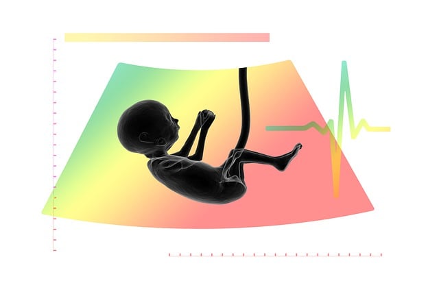
Ultrasound imaging is a non-invasive technique that uses high-frequency sound waves to visualize internal body structures in real-time. It’s a safe and widely accessible method, particularly useful for evaluating organs and tissues during pregnancy, as well as guiding procedures like needle biopsies. However, traditional ultrasound has limitations when it comes to distinguishing between different types of soft tissue, especially small or deeply located tumors. This is where ultrasound imaging with contrast plays a pivotal role.
Contrast agents, when injected into the bloodstream, can enhance the visual distinction between normal and abnormal tissues in ultrasound images. By improving the contrast, these agents help radiologists better detect subtle anomalies, including small tumors that might be missed by conventional ultrasound. This advanced technique significantly improves the accuracy and sensitivity of tumor detection, making it a valuable tool in the early diagnosis and assessment of various conditions.
The Role of Contrast Enhancement in Tumor Detection
Contrast-enhanced ultrasound (CEU) plays a pivotal role in tumor detection, significantly enhancing the sensitivity and specificity of traditional ultrasound imaging. By introducing contrast agents into the body, CEU allows for improved visualization of blood vessels and tissue structures within the suspected tumor area. These agents act as tiny tags, reflecting sound waves differently than surrounding healthy tissues, thus highlighting abnormal regions.
This technique is particularly valuable in differentiating benign from malignant growths, as contrast agents can reveal microvascular networks unique to tumors. The enhanced imaging provides healthcare professionals with crucial information about a patient’s condition, aiding in precise diagnosis and guiding subsequent treatment plans, ultimately improving clinical outcomes in the management of various types of cancer.
How Contrast-Enhanced Ultrasound Improves Accuracy
Contrast-enhanced ultrasound (CEU) significantly enhances the accuracy of tumor detection compared to standard ultrasound imaging. By introducing a contrasting agent into the patient’s bloodstream, CEU allows for better visualization of blood vessels and tissues. This improves the distinction between normal and abnormal structures, making it easier for sonographers to identify tumors that might be obscured or hard to differentiate in standard ultrasound images.
The use of contrast agents in ultrasound imaging provides real-time, high-resolution details about tumor vascularity, size, and shape. This added information helps radiologists interpret the findings more accurately, leading to earlier and more precise diagnoses. As a result, CEU plays a crucial role in improving patient outcomes and guiding effective treatment strategies for various types of tumors.
Benefits and Future Perspectives of CEUS in Oncology
Contrast-enhanced ultrasound (CEUS) offers several significant advantages in tumor detection and management, setting a new standard in oncological diagnostics. By injecting contrast agents into the patient, CEUS allows for real-time visualization of blood flow and tissue structures, enhancing the accuracy of lesion identification and characterization. This non-invasive technique provides detailed information about tumor vascularity, size, shape, and margins, enabling more precise staging and planning of therapeutic interventions.
Looking ahead, the future of CEUS in oncology appears promising. Ongoing research focuses on developing advanced contrast agents with improved targeting capabilities and longer retention times, further refining image quality and diagnostic accuracy. Integrating CEUS into multidisciplinary care teams can facilitate early detection, personalized treatment strategies, and better clinical outcomes for patients. As technology advances, CEUS is poised to become an indispensable tool in the ongoing battle against cancer, offering a safe, accessible, and cost-effective alternative to more invasive imaging modalities.
Contrast-enhanced ultrasound (CEUS) offers a valuable tool for tumor detection, overcoming some limitations of traditional ultrasound. By enhancing visual clarity and distinction between healthy and abnormal tissue, CEUS improves diagnostic accuracy in various oncological settings. This non-invasive technique has proven beneficial in real-world applications, and ongoing research continues to explore its potential in the future of oncology, potentially revolutionizing tumor monitoring and treatment planning.